Rotator Cuff Tendinitis
The shoulder joint has a large range of multi directional movement. Joints that are highly mobile tend to be more unstable. To achieve a high mobility the shoulder depends on dynamic stability from the rotator cuff muscles rather than static stability from ligaments. Tendons around the shoulder joint form the ‘rotator cuff’

Normal shoulder function also depends on the position + movement of the shoulder blade, swelling of the AC joint and the shape / orientation of the acromion (bony point of the shoulder). They also impact on the risk of developing tendonitis or impingement at the shoulder.
Tendonitis of the rotator cuff is a common cause of shoulder pain. It is frequently due to repetitive overuse or over exertion of the attached muscle. It can occur as a secondary issue to poor slouched posture in older people. Repeated use of the arm at or above shoulder level, can provoke inflammation. A fall onto the hand or elbow or a sudden drag on the shoulder / arm can also provoke acute symptoms.


Impingement of the tendon can occur at the shoulder between the head of the humerus and the acromion during vigorous, repetitive activity at or above shoulder level - movements that combine forward flexion to 90' and inward rotation of the arm - as occurs in swimmers, throwers, racquet players.
In older people there are changes within the acromion, the ligaments around the shoulder and the position of the scapula which increase the risk of impingement. The tendons can become inflamed, swell and sore. If this becomes chronic it can cause scarring, weakness and then tearing.
This condition may lead to significantly reduced movement and pain, ultimately causing ‘frozen shoulder’. It responds slowly but well to conservative treatment including exercise and joint mobilisation. Increased strength + flexibility of the muscles around the shoulder and postural retraining all help to improve things in the longer term.
For further information and an assessment please contact us at the clinic.
The following video shows some possible exercises that may be suitable for someone with shoulder + rotator cuff problems. An alternative video is at this link. Specific advice should be sought from your chartered physiotherapist.
Shoulder Dislocation

There are different types of dislocation
-
95% are forward - usually caused by a direct blow or a fall backwards or sideways onto an outstretched arm as above.
-
Backward dislocations occur due to seizure or by strength imbalance of the rotator cuff muscles.
-
Downward dislocation is very unlikely (<1%).
Signs and symptoms
-
Significant pain
-
Inability to move the arm
-
Numbness of the arm
-
Visibly displaced shoulder / flattened appearance around the shoulder.
-
Trauma during dislocation disturbs normal activity of shoulder muscles
-
First-time traumatic dislocation have a high rate of labral injury

Treatments
-
Prompt medical treatment should be sought for any dislocation injury to return the shoulder to its normal position.
-
x-ray will confirm the joint has been replaced into the joint.
-
Rest in a sling for several days until pain subsides
-
Mobilise / exercise the joint as soon as the pain allows.
-
Rotator cuff and deltoid muscle strengthening.
-
Surgery may be required following repeated dislocation.
-
Return to full function and early return to sport or work can be facilitated following treatment and advice from Ennis Physiotherapy Clinic
Shoulder dislocation is a common traumatic injury which can occur during sporting activity or simply as a result of a fall.
A dislocated shoulder occurs when the head of the arm bone separates from the shoulder socket in the shoulder blade. The shoulder has great mobility but little stability and is susceptible to dislocation + subluxation - a partial dislocation.
Tennis / Golfers Elbow
‘Tennis elbow’ is a generic term used to describe a tendinopathy which is an overuse injury of the tendons at the outside of the elbow. It may also be called lateral epicondylitis or as a tendinitis (acute) or tendinosis (chronic) injury.
‘Golfers elbow’ is a similar condition affecting the tendons on the inside of the elbow and is also called medial epicondylitis.
Neither is from playing tennis or golf, but more with the excessive use of the muscles attached to the bone at the elbow - the wrist extensors affecting the outside and the flexors affecting the inside of the elbow..
It occurs where there is excessive repetitive activity, gripping or working with the wrist + hand in an awkward / poor ergonomic position.
Epicondylitis or tendonitis around the elbow responds well to treatment by physiotherapists.
Assessment will identify the key factors are that led to the development of the condition.
Treatment may include:
Avoid the provoking activity for a short period
Soft tissue massage
Stretching + strengthening exercise for affected muscles.
Adjusting work and playing postures / sports equipment.
Electrotherapy Modalities
Medications - prescribed by a GP
A support brace to support the affected tendon to allows ‘rest’.
For an assessment or treatment contact Ennis Physiotherapy Clinic.
Wrist / Colles Fracture
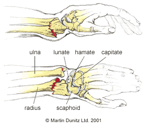
A broken wrist can occur after a fall onto an outstretched hand. The break occurs at the lower end of the bone(s) of the forearm, usually the radius bone. The bone tilts backwards in a Colles Fracture. If the bone fragment tilts forward it is known as a Smith’s fracture.
There is usually distortion of the shape of the wrist, severe pain and minimal active movement is possible. The area can become bruised and swollen within a short period and it can extend into the hand + fingers.


Treatment
-
Reduction (re-alignment) of the bones + Immobilisation in a cast for a number of weeks..
-
Rehab with active use of the wrist + hand.
-
Gradual return to normal function.
-
Physiotherapy where there is difficulty with returning to full range of movement or in restoring normal strength and dexterity.
At Ennis Physiotherapy Clinic we can provide detailed assessment and personalised treatment to maximising normal movement in the wrist + hand, minimising swelling + pain, facilitate return to ADL, work, + leisure activities
Fractured Knuckle
Fracture of the knuckles usually occur due to direct impact of rounded lower end of the metatarsal bone, which forms the knuckle of the hand, against hard surfaces.
The middle knuckles is usually slightly prominent in a closed fist so is at highest risk of fracturing. .
An open hand can be struck directly which can also cause a fracture too.
There may be pain, bruising and swelling and reduced movement.
The knuckle may seem to be become more prominent or disappear if the broken fragment moves.


Physiotherapy can help to restore normal movement to the joints of the affected fingers and dexterity in the hand in general.
For treatment and advice or an assessment contact us at Ennis Physiotherapy Clinic


